- Call Now (905) 332-9660
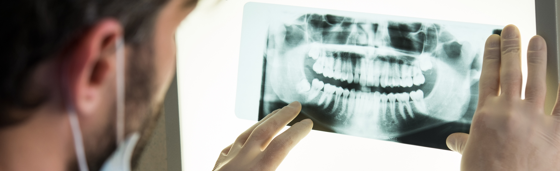
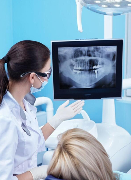
Radiographs, sometimes known as dental X-rays, are interior pictures of your jaws and teeth. Dentists utilize X-rays to look at structures such as your jawbone, nerves, sinuses, and tooth roots that are not visible during a standard examination.
Dental X-rays utilize electromagnetic radiation to create pictures of your mouth, much like X-rays taken of other regions of your body. Your teeth and bones are imaged by the radiation beam as it travels through your soft tissues.
Dental X-rays may be taken digitally, using a computer and digital sensors, or conventionally, using film. Comparing digital dental X-rays to conventional dental X-ray devices, the former need 80% to 90% less radiation.
Dental X-rays aid in the diagnosis of many different oral health conditions by your dentist.
Dental X-rays reveal:
X-rays are also used by dentists to assess your suitability for orthodontics, dentures, dental implants, and other procedures. Additionally, your dentist uses X-rays to monitor the healing process after specific operations including root canal therapy and dental bone grafting. For more information on dental x-rays, contact Iris Dental Group in Burlington today.
© Copyright Iris Dental Group All Rights Reserved.